This 1.5-year-old male baby was referred in February 1994 with multiple anatomic problems depicted below in the Before figure. Cutaneous vesicostomy had been performed elsewhere as an infant. Previous studies had shown a horseshoe kidney, the left side of which was dysplastic. The right ureter ended in a retrovesical “cyst.” Because of vesicoureteral reflux, he had been maintained since infancy on prophylactic antibiotics. Prolonged endoscopy, through duplex urethras, and the vesicostomy, plus appropriate radiology studies, disclosed the anatomy seen in the Before figure.
In a 10-hour operation the dysplastic right kidney was removed, together with the retro vesical “cyst” formed by the lower end of the right ureter, and the right seminal vesicle and vas. The right ureter was reimplanted, repairing the adjacent diverticulum as well. The perineal sinus tract connected with the prostatic urethra was excised. The septum between the two urethras was incised endoscopically.
At age 3 years, endoscopic cutting of the septum between his two penile urethras was performed, creating a single urethral meatus, and correcting his mild hypospadias.
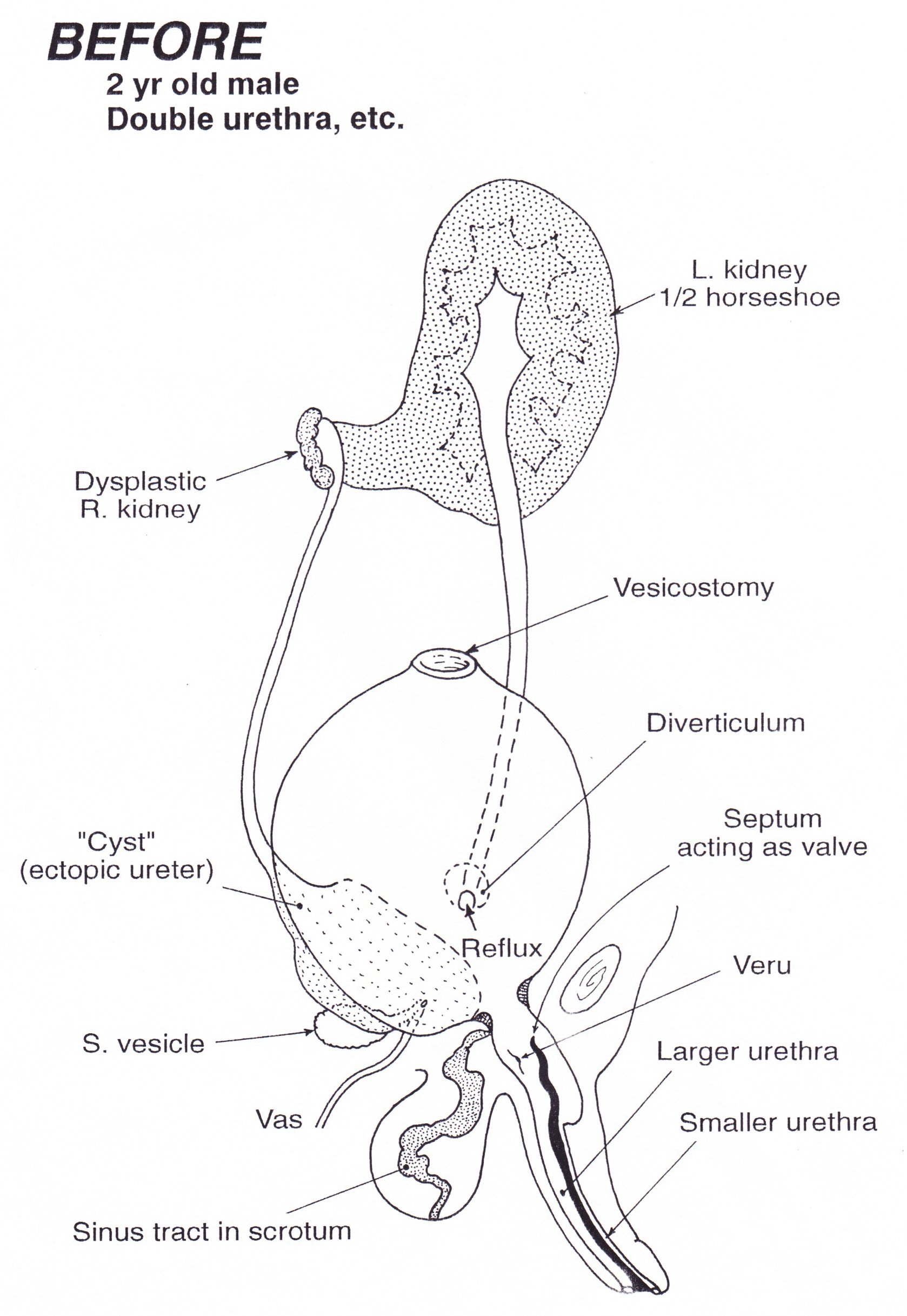
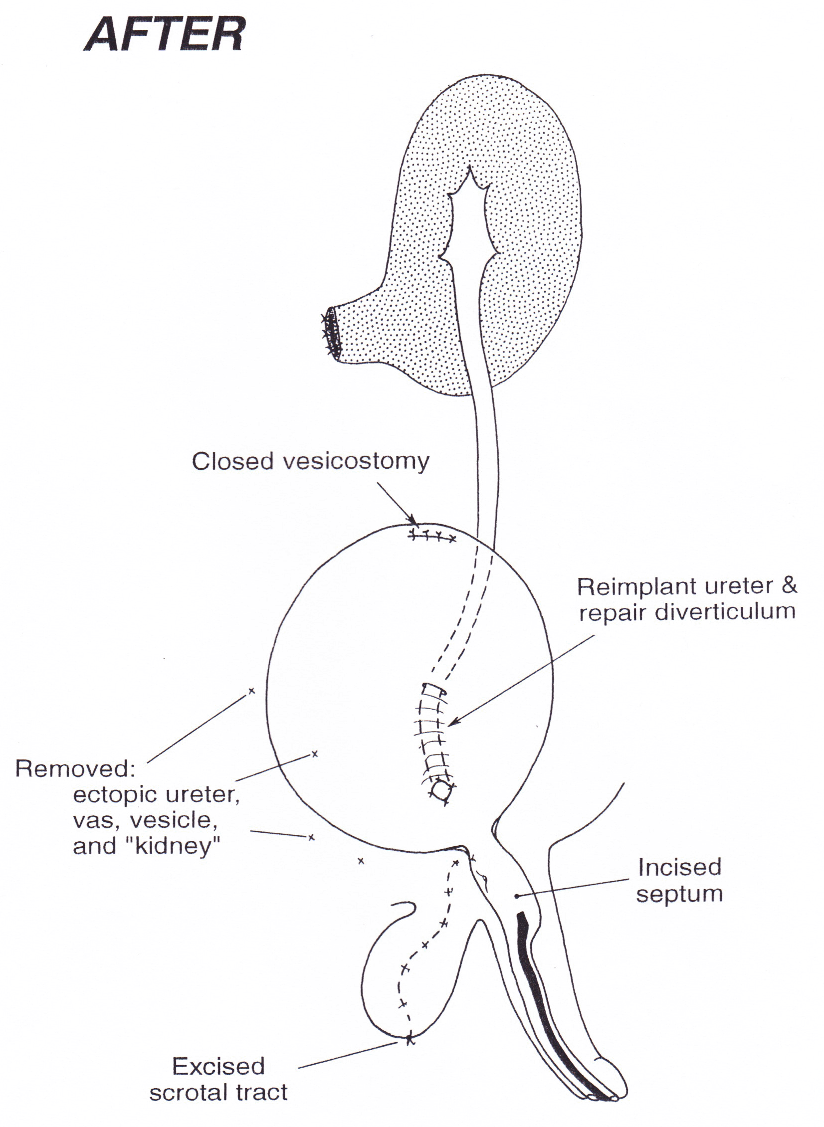
Anatomy (Select Image for High-quality Version). (Top) Before undiversion. (Bottom) After undiversion.
The patient developed well physically (height 6’5”). He is continent of urine and sexually active.