Early Esophageal Atresia Managed by Closure of the Tracheoesophageal Fistula (Hendren Patient Case)
One of the early survivors among 12 babies with esophageal atresia was managed by closure of the tracheoesophageal fistula (TEF), gastrostomy, and exteriorization of the cervical esophagus to the left neck as a neonate. Then a subcutaneous presternal “esophagus” was made in stages.
 n
n
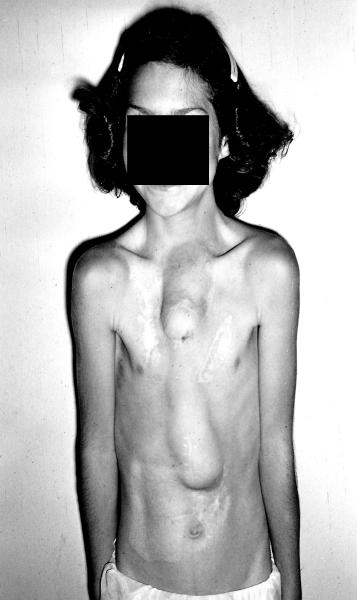
- 11-yr-old female patient born in 1944. Her first surgery was done by William Ladd, M.D., and Orvar Swenson, M.D. It included the above as an eonate plus repair of duodenal atresia. Upper presternal tube is a (skin-graft-lined) skin flap tube connected to the cervical esophagus. Lower half of the “esophagus” was a roux-en-y jejunal tube. Her swallowed food was manually massaged downward.
- In these early cases, the stomach was bypassed. They grew poorly until the stomach was functionalized.
- This child had 30 prior operations in her lifetime. I met her at age 16. A substernal colonic esophagus was constructed using left colon, antiperistaltically. The original subcutaneous tube was moved later.
- At age 19 years, large secundum atrial septal defect was closed using a dacron patch during cardiopulmonary bypass. Substernal colon was carefully removed aside to access the heart.