Tracheoesophageal Fistula

20-Year-Old Female Patient with Esophageal Atresia with Tracheoesophageal Fistula Repaired at Birth (Hendren Patient Case)

  • 20-year-old female had esophageal atresia with tracheoesophageal fistula (TEF) successfully repaired at birth in a nearby hospital.
  • At age 17 years, we saw her for a “cold nodule” of the left thyroid.
  • Hemithyroidectomy was performed for what proved to be a follicular adenoma
  • Barium swallow at that age showed dysmotility of the esophagus commonly seen following repair of esophageal atresia, but not gastroesophageal reflux or other abnormality.
  • She now had a history of dysphagia for 3 mo when swallowing solid foods, and a weight loss of 4.5 kg (10 pounds).
  • Barium swallow showed an intraluminal mass at the esophagogastric junction.

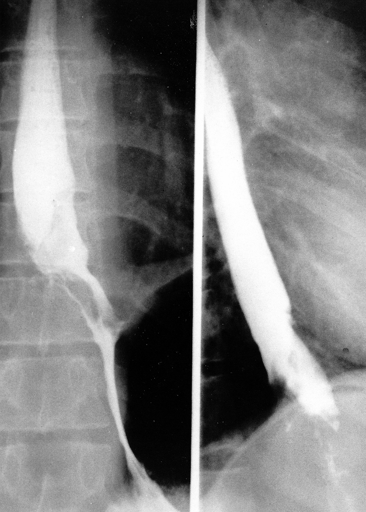
Fig. 1. Pre-Operative Barium Swallow (Select Image for High-quality Version). This demonstrates an irregular intraluminal lesion at and above the gastroesophageal junction.

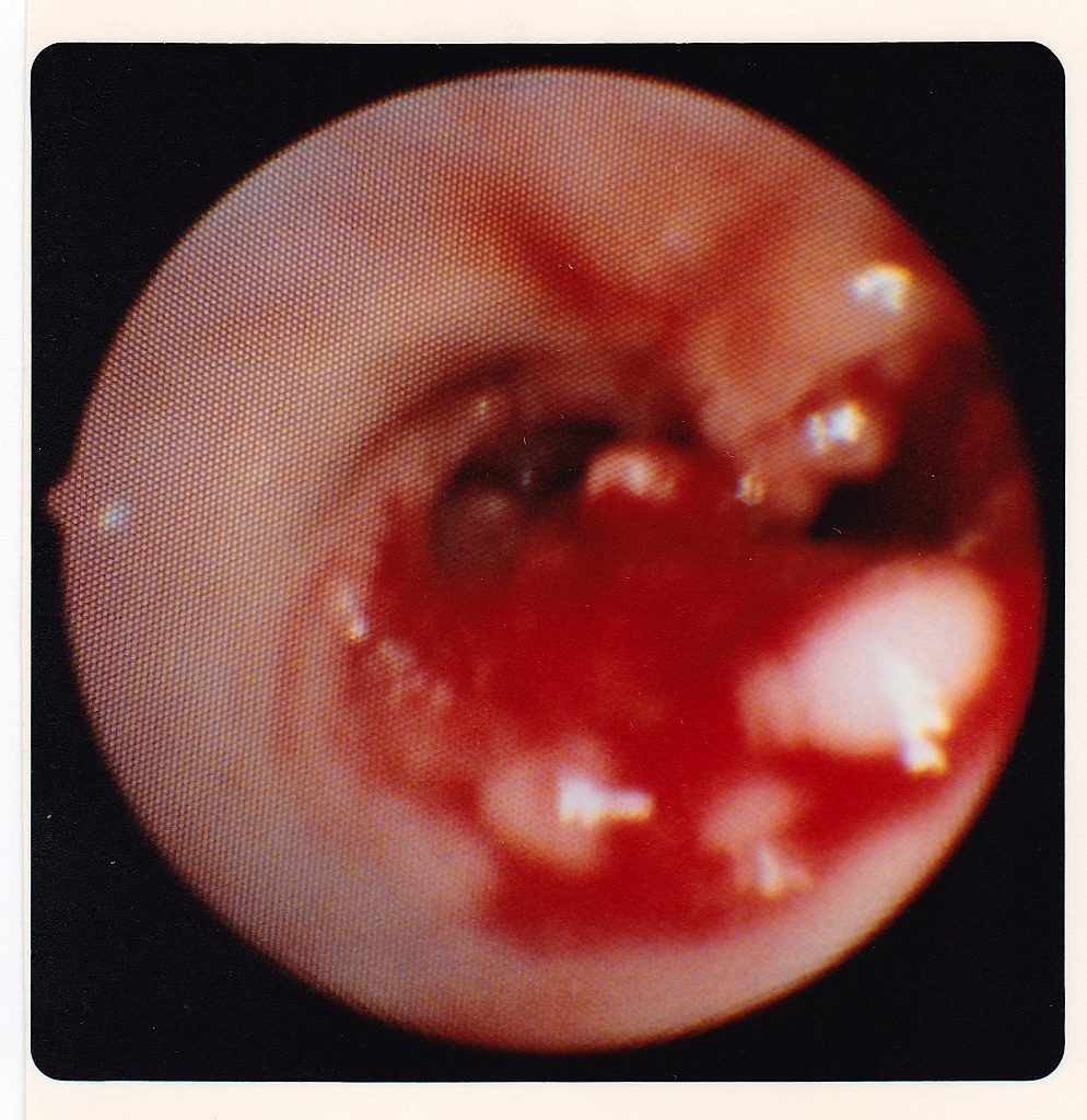
Fig. 2. Endoscopic View of Upper End of Esophagogastric Adenocarcinoma (Select Image for High-quality Version). Esophagoscopy showed an obvious carcinoma of the esophagus, a well differentiated adenocarcinoma histologically.

Treatment Strategy

  • Using a left thoracoabdominal approach, tumor was removed and all lymph nodes in the specimen were negative microscopically for tumor.
  • Patient had an uneventful recovery from the esophagogastrectomy in November 1987.

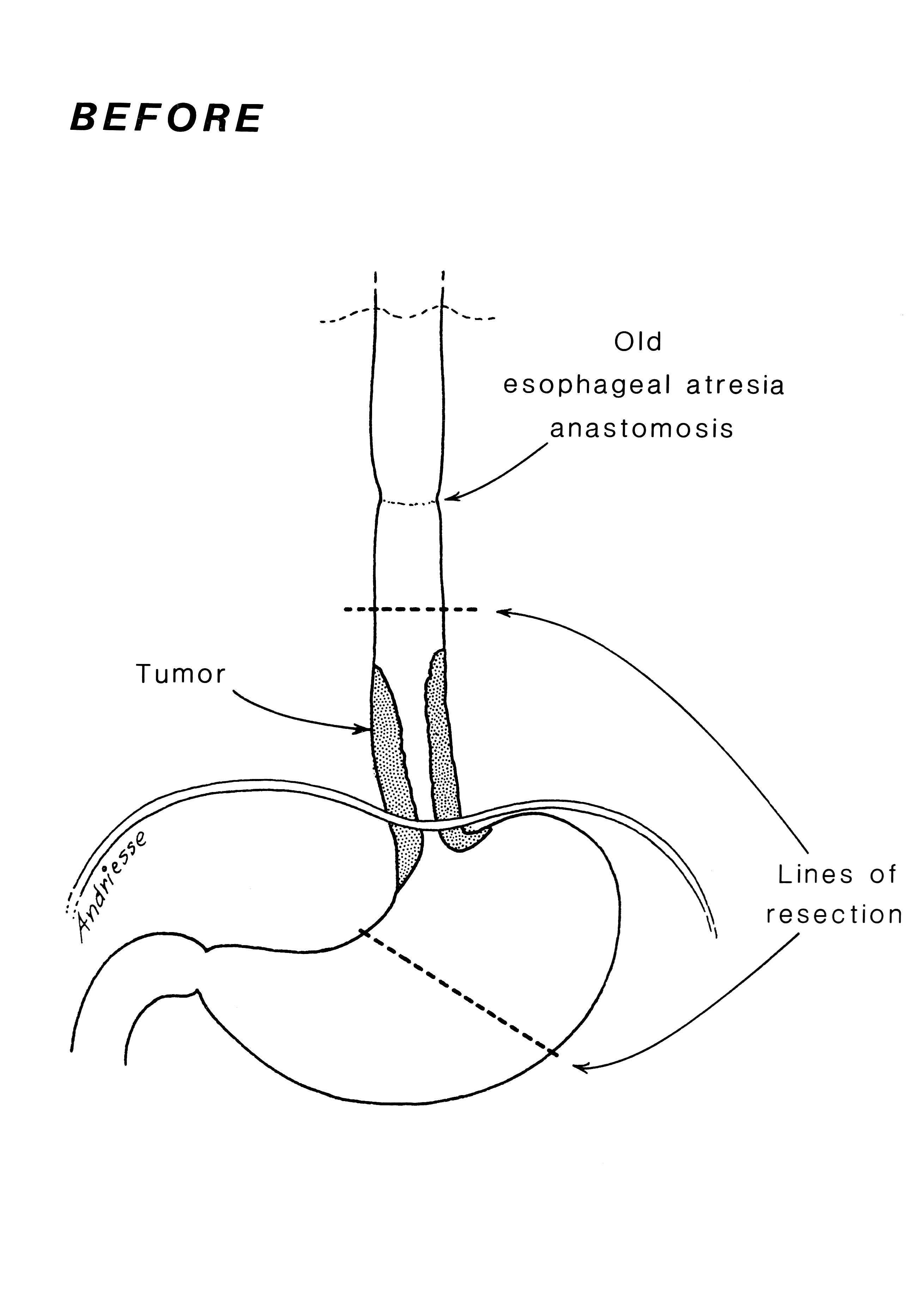
Fig. 3. Pre-Operative Anatomy (Select Image for High-quality Version). Diagram shows the extent of tumor and of resection to be performed using a thoracoabdominal exposure. 

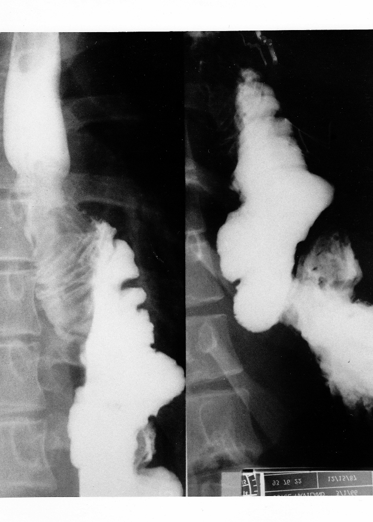
Fig. 4. Post-Operative Barium Swallow (Select Image for High-quality Version). This shows colon interposition from mid esophagus to remaining stomach.

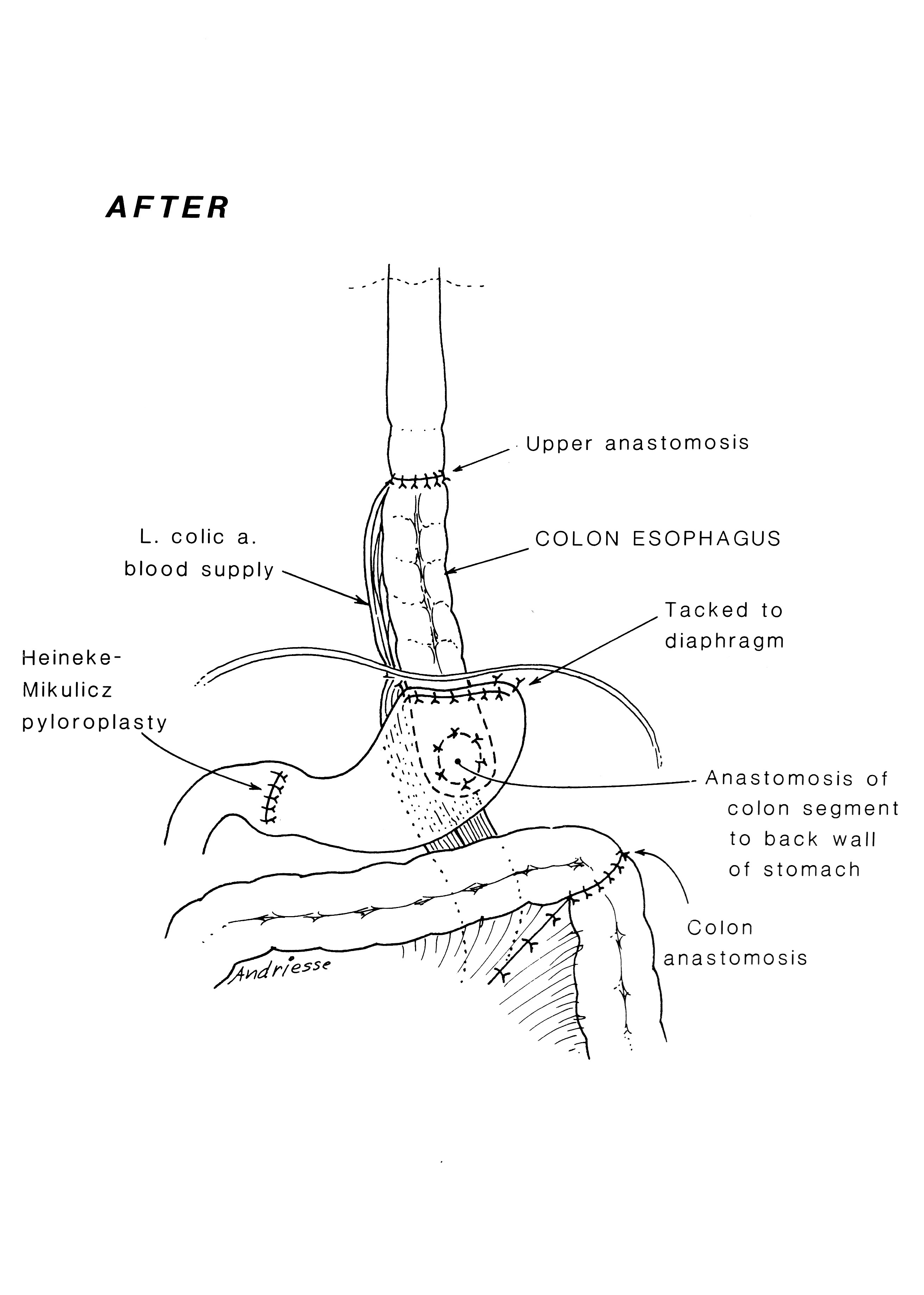
Fig. 5. Post-Operative Anatomy (Select Image for High-quality Version). Anatomy after tumor resection, and restoring gastrointestinal continuity with segment of transverse colon on left gastric pedicle. Note pyloroplasty to enhance gastric emptying.

Follow Up

  • In 1992, 15 years later at age 26 years we performed a left thyroid gland total removal.
  • Histology showed benign modular goiter.
  • Patient last seen in 1994 and she was well.

2-Year-Old Female Patient from Guatemala Born with Esophageal Atresia and Tracheoesophageal Fistula (Hendren Patient Case)

  • 2-year-old female patient from Guatemala born with esophageal atresia and tracheoesophageal fistula (TEF).
  • She had been treated by division and closure of TEF, gastrostomy, and marsupialization of the upper esophagus into the left neck.
  • She was referred to Richard H. Sweet, M.D., at Massachusetts General Hospital in October 1953. Dr. Sweet brought her stomach to her neck to restore her gastrointestinal continuity. It was my privilege to be a surgical resident on Dr. Sweet’s service at that time.
  • By chance, 26 years later, she was sent to me for symptoms of severe gastritis, in her own words, “like a volcano in my chest,” and fullness after eating with respiratory distress by compression of her left lung when her stomach was full.
  • Her stomach emptied very slowly on Barium examination. Pyloroplasty had not been done when she was a baby.

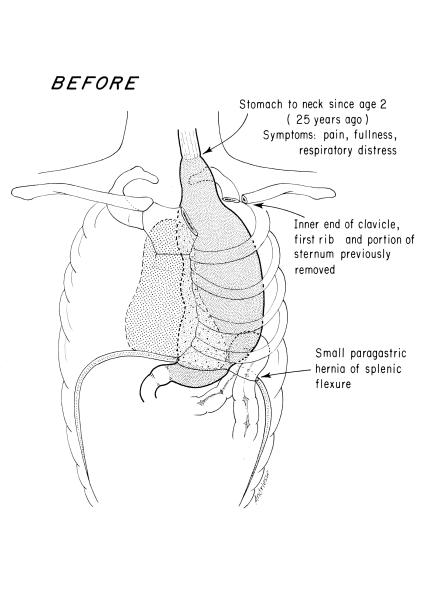
Fig. 1. Pre-Operative Anatomy. Gastric “pull-up operation” done in childhood. 

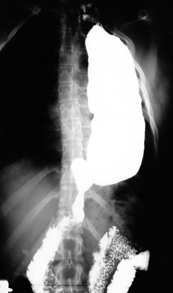
Fig. 2. Barium Filled Stomach. Upper G.I. series age 28 years, with severe symptoms secondary to stomach in the thorax.

Treatment Strategy

  • In a 13-hour thoracoabdominal and left neck procedure in November 1979, the stomach was mobilized and placed back in the abdomen.
  • Generous pyloroplasty was performed.
  • Colon esophagus was fashioned from the transverse colon, anastamosing its hepatic flexure end-to-end with the cervical esophagus and its splenic flexure to the back wall of the stomach.
  • Blood supply of the conduit was left colic and left half of the mid colic artery.
  • Right gastroepiploic vessel was carefully spared, being the principal vessel perfusing the stomach. 

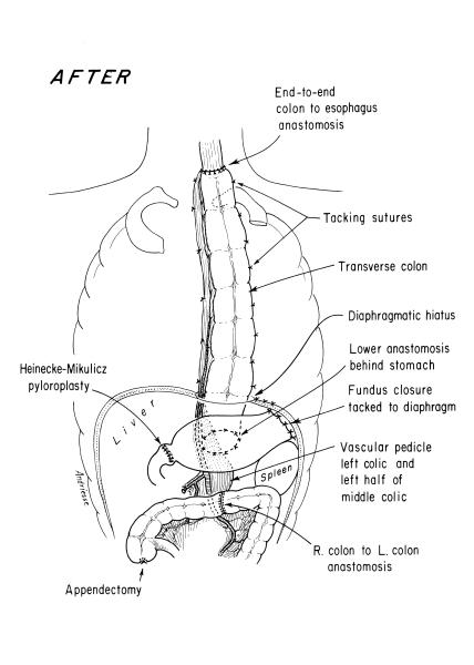
Fig. 3. Post-Operative Anatomy. Esophagoscopy immediately preceding the surgery interestingly showed no esophagitis in the segment just proximal to the gastric fundus in the upper thorax. The scope could pass easily all the way into the duodenum without encountering inflammatory changes.

Follow Up

  • Patient convalesced uneventfully and returned to Guatemala, where we had the pleasure of visiting her the following year. She was asymptomatic.
  • In 1984, she returned to Boston with symptoms of epigastric discomfort 2 hours after meals.
  • Various medications were used, with only transient relief.
  • Gastroscopy by an experienced gastroenterologist disclosed no cause for symptoms.
  • Infusion of acid, saline, alkali, or her gastric juice did not cause her symptoms.
  • Sucralfate was prescribed.
  • Patient last seen in 1997, at age 46 years, by a second, very experienced gastroenterologist.
  • Endoscopy of the colon esophagus and the stomach were unremarkable.
  • No additional contact.

Early Esophageal Atresia Managed by Closure of the Tracheoesophageal Fistula (Hendren Patient Case)

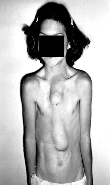
One of the early survivors among 12 babies with esophageal atresia was managed by closure of the tracheoesophageal fistula (TEF), gastrostomy, and exteriorization of the cervical esophagus to the left neck as a neonate. Then a subcutaneous presternal “esophagus” was made in stages.

n

  • 11-yr-old female patient born in 1944. Her first surgery was done by William Ladd, M.D., and Orvar Swenson, M.D. It included the above as an eonate plus repair of duodenal atresia. Upper presternal tube is a (skin-graft-lined) skin flap tube connected to the cervical esophagus. Lower half of the “esophagus” was a roux-en-y jejunal tube. Her swallowed food was manually massaged downward.
  • In these early cases, the stomach was bypassed. They grew poorly until the stomach was functionalized.
  • This child had 30 prior operations in her lifetime. I met her at age 16. A substernal colonic esophagus was constructed using left colon, antiperistaltically. The original subcutaneous tube was moved later.
  • At age 19 years, large secundum atrial septal defect was closed using a dacron patch during cardiopulmonary bypass. Substernal colon was carefully removed aside to access the heart.