Illustrative Cases

Newborn Infant Female with Abdominal Distention (Hendren Patient Case)

Newborn infant girl with abdominal distention and no passage of gas or meconium via the urogenital sinus or patent urachus

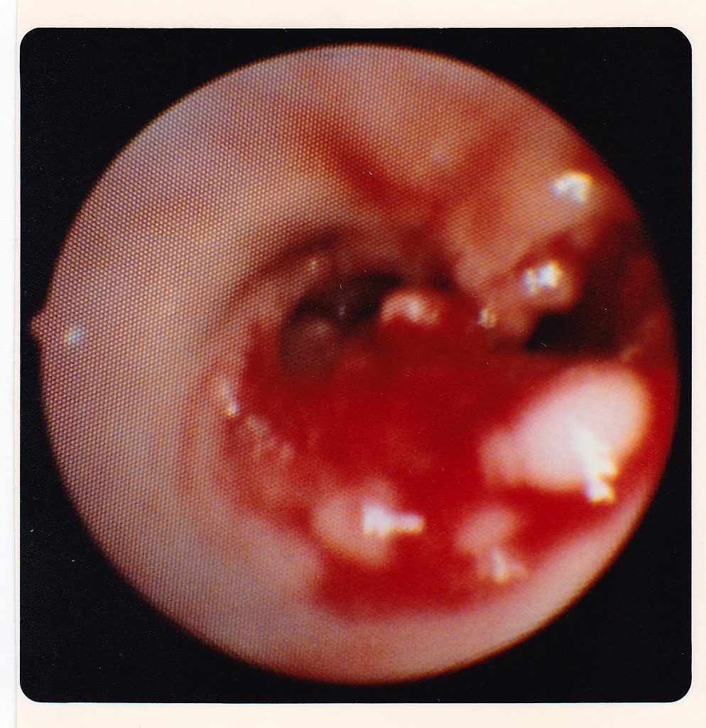
  • Endoscopy and contrast studies unhelpful
  • Laparotomy disclosed ileal atresia and features of cloaca and also cloacal exstrophy
  • Ileostomy and end colostomy at birth
  • 19-hour complete reconstruction at age 23 months

Operative Details

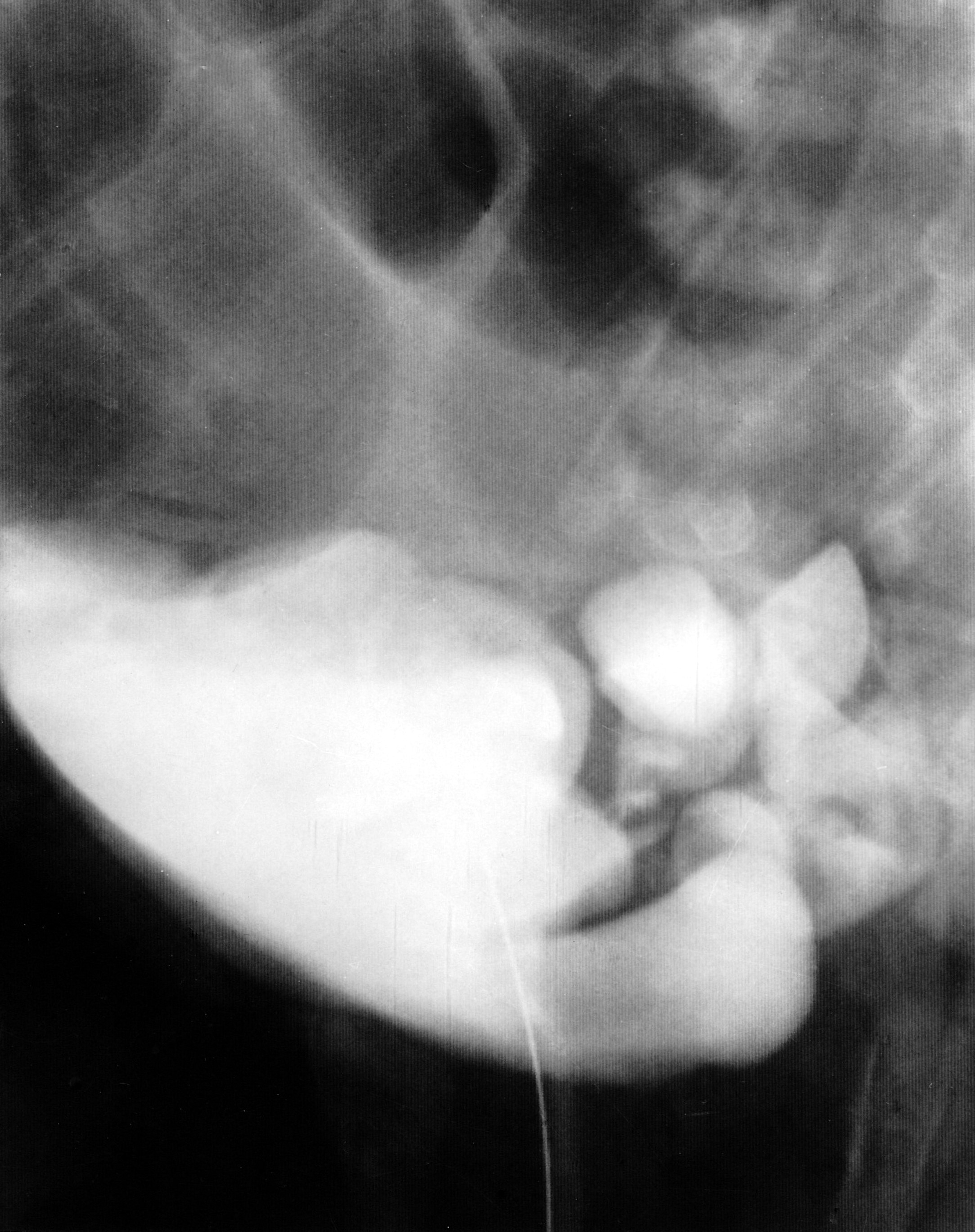
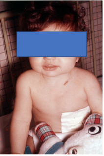
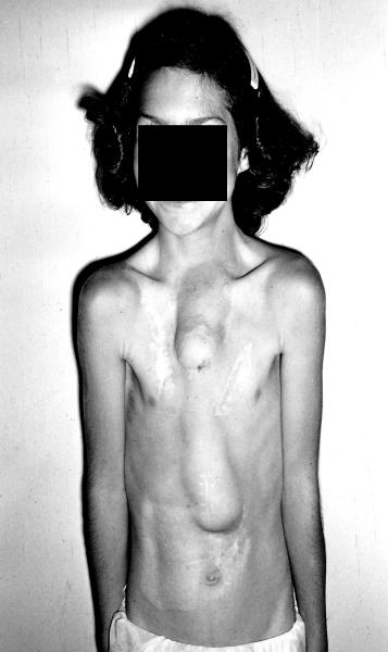
Female patient referred at birth in 1987 with imperforate anus, abdominal distention, and a funnel-shaped appearance to her vaginal introitus (Fig 1). No meconium had passed. Cystogram showed several diverticula arising from the bladder, shedding little light on what the anatomy might be. Endoscopy was performed before laparotomy to relieve bowel obstruction. There was a wide urogenital sinus with no structures entering it. There were multiple openings in the bladder including a patent urachus.

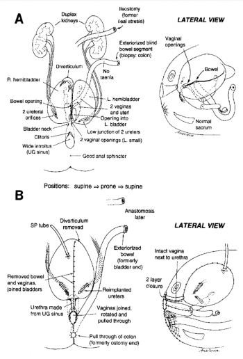
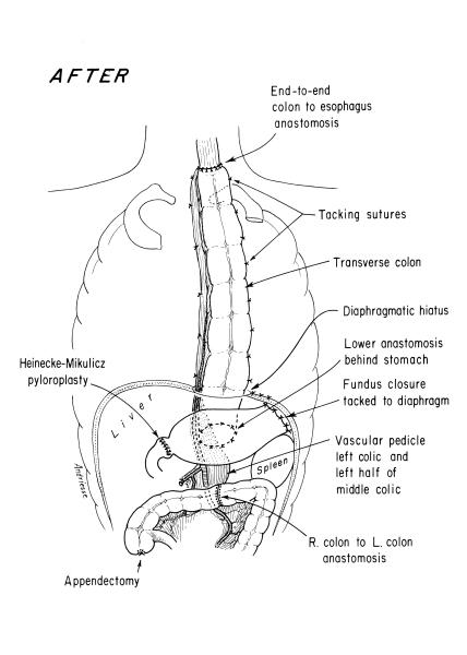
Fig. 1. A. Preoperative anatomy. Two bladders, 2 vaginas and uteri. End ileostomy for ileal atresia at birth, and colostomy for blindly ending microcolon. B. Postoperative anatomy. After cloacal repair at age 2 years, because of its complexity – bladders joined, 4 ureters reimplanted, vaginas joined and rotated to prevent fistula. Note pullthrough of once blind end of microcolon as rectum.

Real atresia was found at laparotomy; below it there was a gap in the mesentery and a short segment of colon in the lower abdomen. The upper end of the colon was blind and the lower end connected with the vagina and bladder which were tethered upward. The right ureter was imbedded in scar surrounding the vaginostomy and required mobilization. The patient was then turned prone. The vagina was separated from the urogenital sinus, which was strictured. The etiology of the stricture appeared to be compression by the pubis because the bladder was pulled upward by the vesicostomy. Resection and reanastomosis of the urogenital sinus was performed. The rectovaginal fistula was divided. The part of the vagina that had been the stoma was brought to the perineum. This was possible because it had been mobilized from above. The rectum was tapered and placed within the muscle complex. The patient was then turned supine to reimplant the right ureter and place a suprapubic tube in the bladder. Endoscopy 2 weeks later showed satisfactory healing. A new urethral catheter was placed to allow further healing. MRI examination showed slight tethering of the spinal cord, but neurosurgical consultation felt that an untethering operation was not needed at that time. Three months later colostomy closure was performed. The patient voided well and intermittent catheterization was discontinued. During the next year constipation was a problem and was relieved by enema washouts. The anal opening became slightly stenotic and was enlarged by 2 z-plasties. At age 4 years, rectosigmoid remained dilated. Sigmoid resection was performed.

Comment: Only case we have seen with an H-fistula from the rectum to the vagina. Furthermore, posterior anal ectopia is extremely rare. When the rectal opening is ectopic, it nearly always lies forward of the point of maximum contractility of the sphincter complex.

The cause for the urogenital sinus stricture may have been created by traumatic catheterization or endoscopy bladder. The urachus was closed. The heal atresia was exteriorized. The blindly ending colon was also exteriorized without knowing whether this was the end of bowel that might have been in continuity with the heal atresia or whether it was the end of bowel as usually seen in cloaca1 exstrophy. At age 7 months endoscopy was performed again, but the findings were difficult to decipher. At age 15 months endoscopy was repeated with uroradiologist, Dr Robert Lebowitz, present with a C-arm fluoroscopic unit. As each orifice was visualized, and catheterized, contrast medium was injected, obtaining fluoroscopy and spot films (Fig 2). The anatomy depicted in Fig 1A was derived in that fashion. There were two hemibladders, four ureters, two vaginas entering the bladders, and a bowel opening in the septum between the two hemibladders. There was a diverticulum at the apex of the right hemibladder with communicating openings.

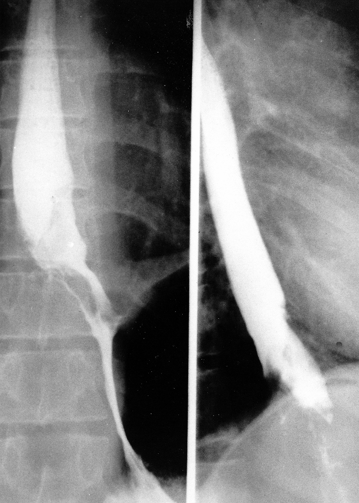
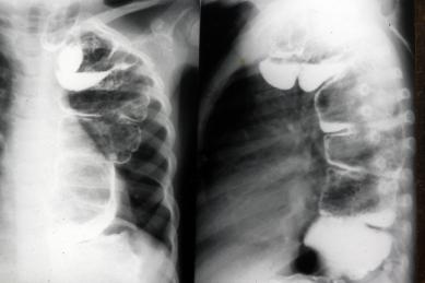
Fig. 2. Preoperative roentgenogram showing bizarre constellation of structures produced by filling anatomy shown in Fig 1A. Prolonged endoscopic study in conjunction with simultaneous roentgen study with C-arm fluoroscopic unit in operating room was required to decipher the anatomy.

Repair was performed (Fig 1B) at age 2 years. After circumferentially preparing and draping the child, a midline laparotomy was performed to dissect out all of the anatomy. Biopsy specimens were taken from both ends of the distal defunctioned bowel segment because it was not possible from the orientation of its mesentery to tell which end was developmentally the distal one, ie, whether the blind end was what might have been in continuity with the ileum, or whether it was the blindly ending gut seen in cloaca1 exstrophy. Interestingly, there were no tinea on the colon. The bladder was opened and it was possible to locate the two vaginas which entered on each side of a midline septum. They were dissected free from the bladder, using the intravesical approach and were sewn together to create a single vagina. The bowel segment was detached from the bladder. The ureters were reimplanted. The midline septum between the bladders was excised to make a single bladder. The patient was then turned into the prone position to make a posterior sagittal midline incision. The wide urogenital sinus was tapered into a urethra and closed with two layers. The two vaginas were rotated 90” and joined to the perineum. A perineal body was constructed. The appropriate end of the bowel segment was pulled through and enclosed in the muscle complex. The child was then turned supine to complete bladder closure and exteriorize the other end of bowel. The anesthesia time for this global reconstruction was 20 hours. The patient convalesced uneventfully. Endoscopy 2 weeks later showed satisfactory healing. Intermittent catheterization was begun and the suprapubic tube was soon removed. Four months later bowel continuity was restored by resecting the ileostomy and joining it to the defunctioned distal bowel. The patient could not empty her bladder completely and was maintained on intermittent catheterization. Surprisingly, at age 3 years she emptied her bowel 2 to 3 times daily voluntarily and without soiling. A minor degree of rectal prolapse was trimmed as an outpatient procedure. MRI of the spine showed no tethering of her spinal cord. She now voids spontaneously, does not wet, and is catheterized for residual twice daily as a precaution, usually obtaining about 100 mL.

Comment: This case is of special interest because the anatomy initially appeared so complex that there seemed little likelihood that reconstruction would achieve continence. Those gloomy prospects proved incorrect. It was vital to thoroughly assess the anatomy both radiographically and endoscopically. This case seems to be a transition form between a usual cloaca and cloacal exstrophy, because it included hemibladders, vesicointestinal fissure, and a blindly ending segment of bowel hanging in the pelvis. However, there was no omphalocele or bladder exstrophy.

Additional Images:

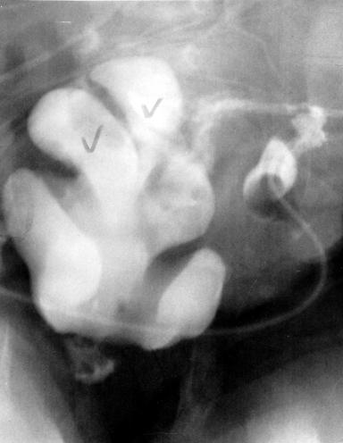
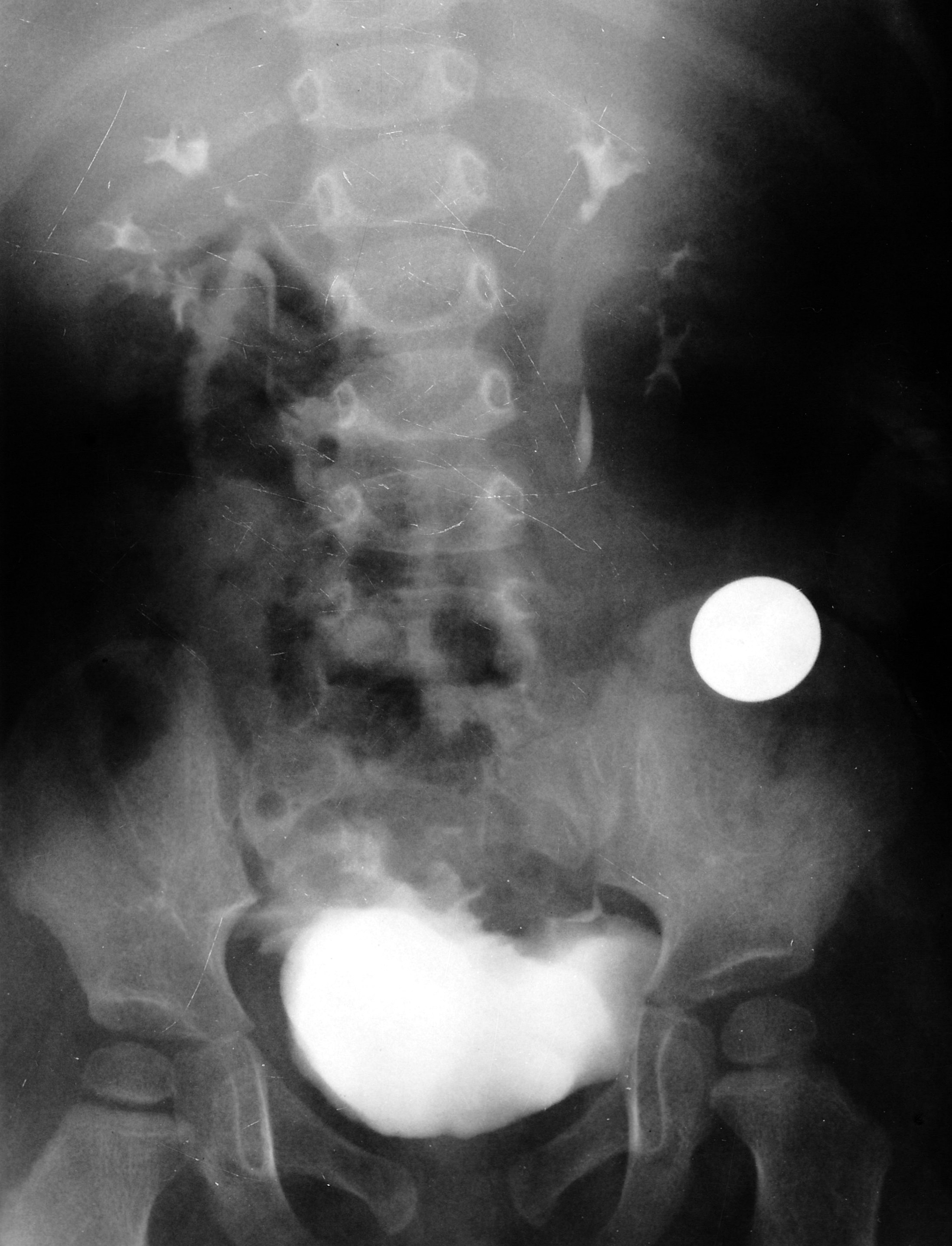
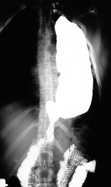
Fig. 3. A.P. View of pelvic structures filled with contrast.  "Undecipherable" by radiologist or surgeon!

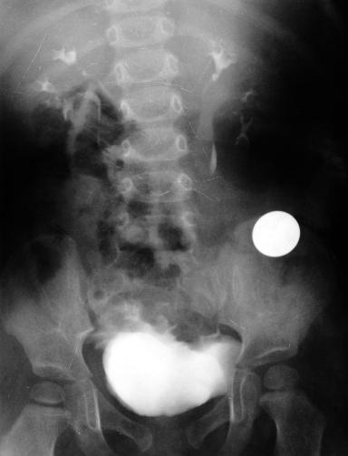
Fig. 4. Note swallowed coin in ileum, removed with grasping forceps before ileo-colic anastamosis 4 months post cloaca repair.  Normal upper tracts and single bladder.


Early Cloacal Reconstruction Case: Evolution of Treament Strategies (Hendren Patient Case)

  • Neonate treated in 1975--early in experience with cloacal reconstruction
  • Sigmoid colostomy was done, but transverse colostomy now preferred
  • Demonstrated repair in lithotomy position to be possible, although today a posterior sagittal approach might be used
  • Hendren's first cloacal repair in 1962 had the vagina exteriorized with a perineal flap, and the rectum was repaired by a posterior sagittal approach and pullthrough

20-Year-Old Female Patient with Esophageal Atresia with Tracheoesophageal Fistula Repaired at Birth (Hendren Patient Case)

  • 20-year-old female had esophageal atresia with tracheoesophageal fistula (TEF) successfully repaired at birth in a nearby hospital.
  • At age 17 years, we saw her for a “cold nodule” of the left thyroid.
  • Hemithyroidectomy was performed for what proved to be a follicular adenoma
  • Barium swallow at that age showed dysmotility of the esophagus commonly seen following repair of esophageal atresia, but not gastroesophageal reflux or other abnormality.
  • She now had a history of dysphagia for 3 mo when swallowing solid foods, and a weight loss of 4.5 kg (10 pounds).
  • Barium swallow showed an intraluminal mass at the esophagogastric junction.

Fig. 1. Pre-Operative Barium Swallow (Select Image for High-quality Version). This demonstrates an irregular intraluminal lesion at and above the gastroesophageal junction.

Fig. 2. Endoscopic View of Upper End of Esophagogastric Adenocarcinoma (Select Image for High-quality Version). Esophagoscopy showed an obvious carcinoma of the esophagus, a well differentiated adenocarcinoma histologically.

Treatment Strategy

  • Using a left thoracoabdominal approach, tumor was removed and all lymph nodes in the specimen were negative microscopically for tumor.
  • Patient had an uneventful recovery from the esophagogastrectomy in November 1987.

Fig. 3. Pre-Operative Anatomy (Select Image for High-quality Version). Diagram shows the extent of tumor and of resection to be performed using a thoracoabdominal exposure. 

Fig. 4. Post-Operative Barium Swallow (Select Image for High-quality Version). This shows colon interposition from mid esophagus to remaining stomach.

Fig. 5. Post-Operative Anatomy (Select Image for High-quality Version). Anatomy after tumor resection, and restoring gastrointestinal continuity with segment of transverse colon on left gastric pedicle. Note pyloroplasty to enhance gastric emptying.

Follow Up

  • In 1992, 15 years later at age 26 years we performed a left thyroid gland total removal.
  • Histology showed benign modular goiter.
  • Patient last seen in 1994 and she was well.

2-Year-Old Female Patient from Guatemala Born with Esophageal Atresia and Tracheoesophageal Fistula (Hendren Patient Case)

  • 2-year-old female patient from Guatemala born with esophageal atresia and tracheoesophageal fistula (TEF).
  • She had been treated by division and closure of TEF, gastrostomy, and marsupialization of the upper esophagus into the left neck.
  • She was referred to Richard H. Sweet, M.D., at Massachusetts General Hospital in October 1953. Dr. Sweet brought her stomach to her neck to restore her gastrointestinal continuity. It was my privilege to be a surgical resident on Dr. Sweet’s service at that time.
  • By chance, 26 years later, she was sent to me for symptoms of severe gastritis, in her own words, “like a volcano in my chest,” and fullness after eating with respiratory distress by compression of her left lung when her stomach was full.
  • Her stomach emptied very slowly on Barium examination. Pyloroplasty had not been done when she was a baby.

Fig. 1. Pre-Operative Anatomy. Gastric “pull-up operation” done in childhood. 

Fig. 2. Barium Filled Stomach. Upper G.I. series age 28 years, with severe symptoms secondary to stomach in the thorax.

Treatment Strategy

  • In a 13-hour thoracoabdominal and left neck procedure in November 1979, the stomach was mobilized and placed back in the abdomen.
  • Generous pyloroplasty was performed.
  • Colon esophagus was fashioned from the transverse colon, anastamosing its hepatic flexure end-to-end with the cervical esophagus and its splenic flexure to the back wall of the stomach.
  • Blood supply of the conduit was left colic and left half of the mid colic artery.
  • Right gastroepiploic vessel was carefully spared, being the principal vessel perfusing the stomach. 

Fig. 3. Post-Operative Anatomy. Esophagoscopy immediately preceding the surgery interestingly showed no esophagitis in the segment just proximal to the gastric fundus in the upper thorax. The scope could pass easily all the way into the duodenum without encountering inflammatory changes.

Follow Up

  • Patient convalesced uneventfully and returned to Guatemala, where we had the pleasure of visiting her the following year. She was asymptomatic.
  • In 1984, she returned to Boston with symptoms of epigastric discomfort 2 hours after meals.
  • Various medications were used, with only transient relief.
  • Gastroscopy by an experienced gastroenterologist disclosed no cause for symptoms.
  • Infusion of acid, saline, alkali, or her gastric juice did not cause her symptoms.
  • Sucralfate was prescribed.
  • Patient last seen in 1997, at age 46 years, by a second, very experienced gastroenterologist.
  • Endoscopy of the colon esophagus and the stomach were unremarkable.
  • No additional contact.

Five-Day-Old Female Patient Referred with Esophageal Atresia (Hendren Patient Case)

  • 5-d-old baby girl referred July 1963 with esophageal atresia.
  • X-ray study had shown a blindly ending upper esophagus; abdominal film showed no gas in the abdomen.
  • Stamm gastrostomy using a mushroom catheter was performed, bringing the tube out through a small stab wound.
  • Right chest was opened to confirm for certain that the distal esophagus was rudimentary (having seen a prior case where a small fistula had been present, with a good lower esophagus but without abdominal gas). (Modern imaging would obviate that need.)
  • Upper pouch was mobilized, during which a fistula was seen from the back of the upper pouch to the trachea, not seen on pre-op study.
  • Fistula was divided and closed.
  • After chest closure, infant was turned and a transverse incision in the lower neck just above the clavicle anterior to the sternocleidomastoid muscle and the jugular vein and carotid artery.
  • Previously freed upper esophagus was quickly opened and sewn to the wound to provide free drainage of saliva.
  • Baby thrived.

Fig. 1. Patient at Age 10 Mo Before Colon Esophagus Operation. Note excellent nutritional status. Upper esophagus stoma to drain saliva above left clavicle. Gastrostomy tube covered with ample tape to avoid its being pulled out.

  • At 10 mo, through a left 7th interspace thoracoabdominal incision, a colon esophagus was made using transverse colon on a left colic arterial pedicle (see video of operation).
  • Lower end of colon esophagus did not lie comfortably for anastomosis to the distal esophageal remnant and so it was anastomosed to the anterior wall of the stomach.
  • Postoperatively, when gastrostomy feedings were begun barium study showed free reflux up the colon conduit and less flow through the pylorus, although both vagi were spared intraoperatively.
  • At 2 weeks postop, laparotomy was performed.
  • Gastrostomy tube site was moved farther proximally in the upper stomach.
  • Generous Heineke-Mikulicz pyloroplasty was performed, incising 1 inch (2.54 cm) into the duodenum and 1.5 inches (3.81 cm) into the stomach, closing transversely. Pyloric tissues were indeed thicker than expected.
  • Colon esophagus remained somewhat dilated as if there were functional partial obstruction at its lower end.
  • Some gastrointestinal blood was observed on multiple occasions, never great in amount.
  • At 10 wk, reoperation was performed to accomplish three tasks:
    • (1) shorten the lower end of the colon esophagus;
    • (2) anastomose it to the original rudimentary lower esophagus, as once considered; and
    • (3) divide both vagus nerves to reduce gastric acidity.  

Fig. 2. Barium Swallow in 1968. Although caliber of colon was still somewhat dilated it emptied well, there was no aspiration, and patient was well nourished. 

  • Patient thrived; family moved to Oklahoma.
  • At age 3 yr, she was seen. She and her barium study were both fine.
  • At age 22 yr, patient was and healthy.
  • Unable to get pregnant, she underwent laparoscopy elsewhere; after lysis of fallopian tube adhesions she had two healthy sons.

Takeaways

Early Esophageal Atresia Managed by Closure of the Tracheoesophageal Fistula (Hendren Patient Case)

One of the early survivors among 12 babies with esophageal atresia was managed by closure of the tracheoesophageal fistula (TEF), gastrostomy, and exteriorization of the cervical esophagus to the left neck as a neonate. Then a subcutaneous presternal “esophagus” was made in stages.

n

  • 11-yr-old female patient born in 1944. Her first surgery was done by William Ladd, M.D., and Orvar Swenson, M.D. It included the above as an eonate plus repair of duodenal atresia. Upper presternal tube is a (skin-graft-lined) skin flap tube connected to the cervical esophagus. Lower half of the “esophagus” was a roux-en-y jejunal tube. Her swallowed food was manually massaged downward.
  • In these early cases, the stomach was bypassed. They grew poorly until the stomach was functionalized.
  • This child had 30 prior operations in her lifetime. I met her at age 16. A substernal colonic esophagus was constructed using left colon, antiperistaltically. The original subcutaneous tube was moved later.
  • At age 19 years, large secundum atrial septal defect was closed using a dacron patch during cardiopulmonary bypass. Substernal colon was carefully removed aside to access the heart.